


The company, headquartered in Sao Paulo, designs 3D printed human organs in their actual dimensions. Replicas allow doctors to know in advance and in great detail the area of the body being studied and treated, allowing for more precise surgical procedures
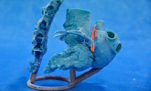
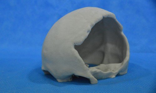
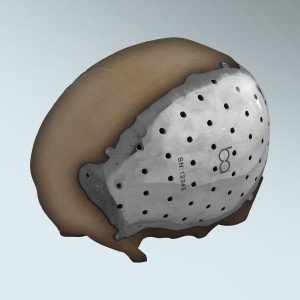
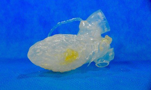
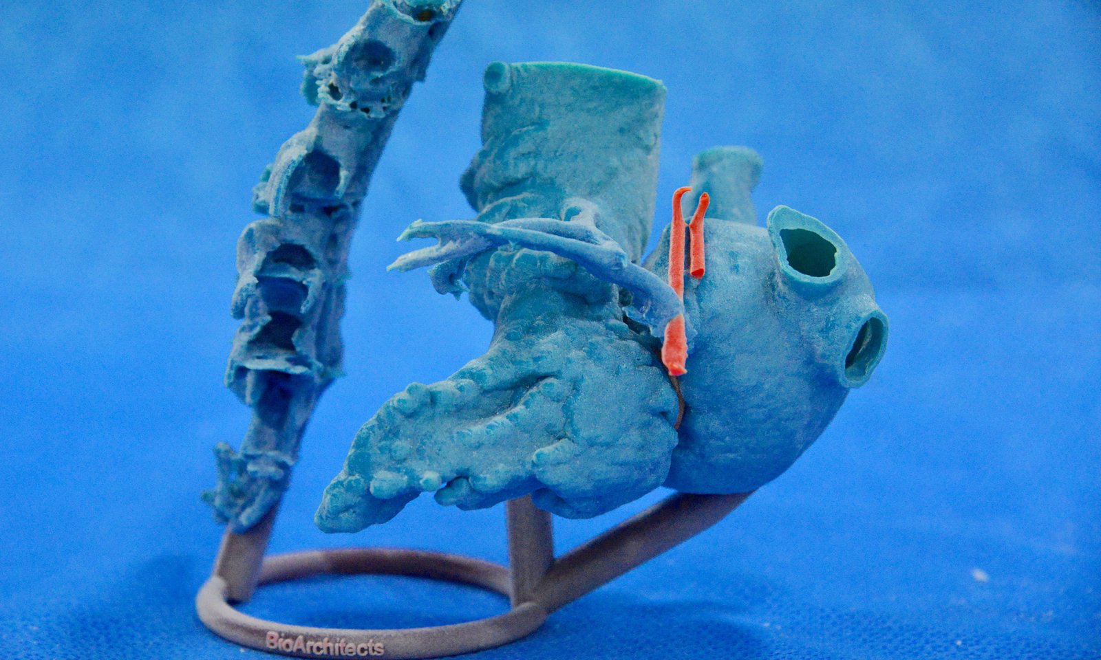
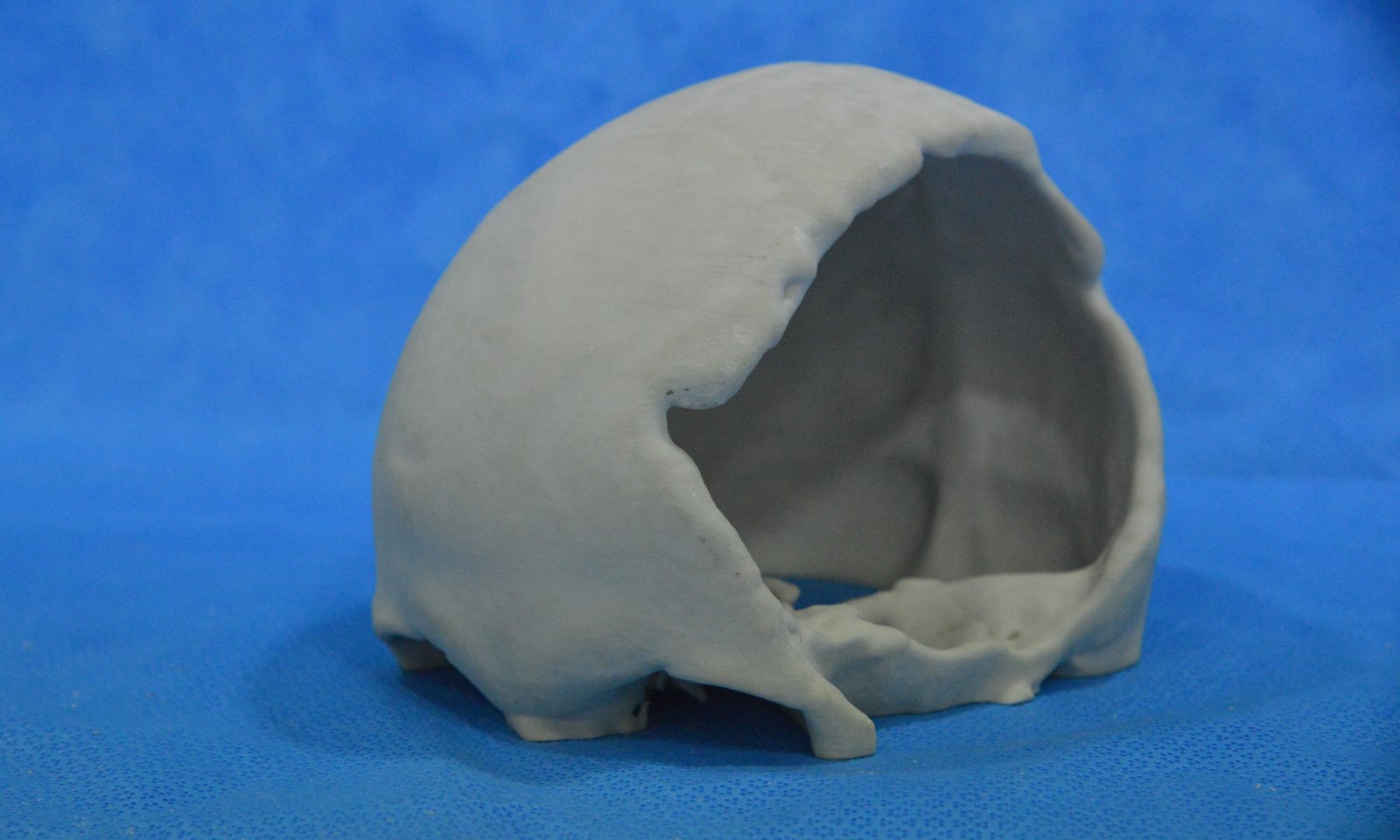
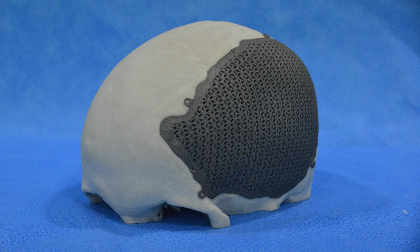
The use of 3D printing in medicine represents one of the greatest scientific advances in recent years. Developing replicas in the true dimensions of human organs allows, among other applications, assistance to the surgeon before an operation. From imaging examinations (such as CT scans, MRIs and PET scans) 3D printers are able to reproduce, in great detail, any part of the body in materials of a diversity of flexibilities, colors and textures. These replicas are called anatomical models. BioArchitects stands out as one of the most innovative Brazilian companies in this market.
“Anatomical models have increasingly become an essential aid for surgical planning, allowing the doctor to ‘practice’ the procedure on a replica before the actual surgery,” explains Walter Troccoli, business development director for BioArchitects, established two years ago, with headquarters in Sao Paulo and also operating in the U.S. since December 2014. Renowned hospitals in Sao Paulo’s capital city have come to know the company’s technology more closely, realizing that it can be a key competitive differentiator and potential tool for reducing costs. Partnerships have been established between BioArchitects and a number of these institutions.
The decision of the company’s partners to invest in the 3D market for medicine emerged from intensive research into which areas of the country showed the most promise for understanding and adopting the full capability of this new technology. Doctors have evidenced curiosity and interest in these advances, influenced as well even by international studies over the past five years highlighting the assistance offered by 3D organ replicas in minimizing risk and bringing a number of health benefits to patients, creating new paradigms in medicine.
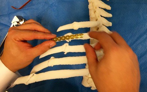
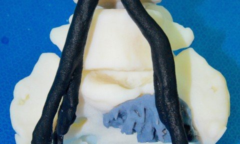
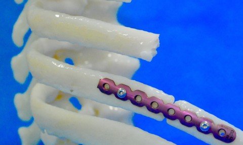
One of BioArchitects’ anatomical models has recently been used for the first time in Brazil, in the surgical repair of multiple fractures of the thorax. The surgeon accurately located the fractures in the ribs of a 65-year-old patient, using titanium plates for their repair. The use of 3D printing technology avoided the waste of materials, and surgical time and resulted in improvements in quality of care. Knowing the necessary corrections in advance, by viewing and practicing on a replica of the patient’s ribs resulted in the use of three titanium plates, instead of the five initially planned for. The anatomical model avoided common surprises, through the discovery of findings such as bones with fractures greater than expected, in addition to assuring the physician surgical access to the exact location of the injury.
Incorporating anatomical models as part of the surgical procedure offered benefits for all involved. For the physician, greater accuracy, better planning and up to a 40% reduction in the time required for the procedure. For the patient, lowered surgical risk, diminished exposure to anesthesia and radiation and a reduced possibility of contamination. “It is also important to emphasize the cost reduction possible through the use of anatomical models,” explained Walter Troccoli, of BioArchitects.
The anatomical model for the planned thoracic surgery was produced on Israeli equipment representing the latest technology. “We brought this machine exclusively for use in Brazil,” said Felipe Marques, COO of the company. “It is a printer capable of producing anatomical models with perfection, printing in multiple materials and a diverse range of colors. This allows us to differentiate between blood vessels, tissue, fat, muscle, etc. No other machine in Brazil can achieve such a level of differentiation. ”
The anatomical models developed by BioArchitects present a wealth of detail that guarantees better results in surgical techniques. The process of providing our service involves several steps, beginning with the imaging examination, the subsequent segmentation of the image for prototyping and finally printing of the model. The time required to complete all these steps varies, but is never more than a few days.


